How the iTrace Works
ADVANCED TECHNOLOGY SHOWS HOW YOUR PATIENTS SEE
WHY RAY TRACING IS ESSENTIAL FOR WAVEFRONT ANALYSIS
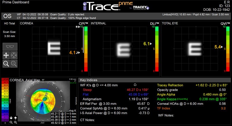
The iTrace is the only device that uses ray tracing for wavefront analysis. It sequentially projects 256 parallel light rays through the entrance of the pupil and detects where each beam of light lands on the retina and how much light energy is transferred. Reviewing this retinal spot pattern point-by-point provides insight into a patient’s overall visual function.
Other devices that rely on a Hartmann-Shack sensor or dynamic skiascopy for wavefront imaging, analyze light as it is reflected back from the retina. Although this works for simple optical systems, these devices are prone to data confusion when measuring highly aberrated eyes, pseudophakia, with cataracts, through contact lenses, or over spectacles.
The iTrace and its proprietary ray tracing technology, however, can directly replicate a patient’s vision under any circumstance.
The Complete Picture: Corneal Topography + Wavefront Aberrometry
Dr. Joe Wakil, CEO of Tracey Technologies, is largely credited with advancing the field of corneal topography. But using a placido disk-based topographic system to map corneal curvature is not what makes the iTrace unique. What differentiates the iTrace is its ability to give insight into both the form and function of the eye.
By integrating wavefront aberrometry with corneal topography, the iTrace can objectively map the internal optics of the eye by subtracting corneal from total aberrations. In under a minute, the iTrace generates a complete profile on a patient’s topography, wavefront, autorefraction, keratometry, day to night vision, pupillometry, white to white and more.
To learn more about how the iTrace functions as a 5-in-1 wavefront aberrometer, corneal topographer, autorefractor, keratometer, and pupillometer, schedule a demo.

TECHNOLOGY DOCTORS TRUST
“The iTrace gives me everything I need. It gives me tremendous wavefront data, it gives me great Placido disk images to look at the quality of my scans, and the Tracey Refraction is probably the most accurate auto-refraction I have available to me.”
BETTER INFORMATION DRIVES BETTER DECISIONS
Objective Evaluation of Poor Vision
Before addressing these visual impairments, you need to identify whether the aberrations are in the cornea or the internal optics. Using the iTrace, practitioners can quickly and accurately identify whether symptoms are caused by the cornea or the lens before recommending a course of treatment.

Lens Dysfunction
This complete set of data allows you to understand exactly where vision issues are coming from, when the right time to recommend a vision correction procedure is, and helps you identify the best method of correction for a specific case.
Premium IOL Candidacy
In addition to looking at this Angle Alpha, the corneal wavefront should also be reviewed. If there are significant corneal aberrations, surgeons can avoid putting a multifocal lens behind what is essentially a multifocal cornea with this quick screening.


Toric Lens Evaluation

Improving Toric Outcomes
Surgeons can improve their precision in toric IOL surgery with the iTrace’s Toric Planner. This integrated calculator utilizes advanced wavefront keratometry readings to derive an amount of cylinder and an axis location.
The iTrace also produces a corresponding external color photograph that can be used to landmark the eye for accurate lens placement during surgery. These eye image overlays are designed to zero out marking error.


Post-Operative Examinations
After a toric implantation, the lens cylinder power should match the corneal steep axis. The iTrace’s wavefront map displays both the axis of the cylinder internally and the corneal steep axis, which is confirmed by topography.
Because ray tracing is the only way to accurately measure through a toric lens, the iTrace is the only device that calculates the predicted post-surgical refraction and automatically suggests any rotation that will optimize the location of a toric lens. A surgeon can easily review the estimated benefits of rotating a toric lens with a patient after understanding how much cylinder is likely to change.
Since the iTrace is the only device that can accurately scan the complex architecture of a premium IOL, these post-surgical benefits are not exclusive to toric lenses. When poor vision occurs after a premium lens implantation, an undilated scan with the iTrace will bring the root cause to light.
Tear Film Analysis
The iTrace includes the Tear Film Index (TFI), which measures Placido ring projections on the surface of the eye and generates an objective performance metric using our proprietary TBUT algorithm.

Want to Learn More?
Request an iTrace Demo
We look forward to giving you an inside look at how the iTrace works. Simply fill in your preferred contact information and a member of the Tracey Technologies team will reach out to schedule your demo.

